Imaging provides you and your doctor with crucial information about your health. With facilities accredited by the American College of Radiology, we provide high-quality imaging and radiology services with some of the most advanced technologies available, empowering you and your doctor with the information you need to make important health care decisions.
A doctor’s ability to diagnose some injuries and illnesses depends on images they have of the affected areas of your body. At CommonSpirit Health, we provide advanced imaging technologies to enable you and your health care team get a better picture of your health.
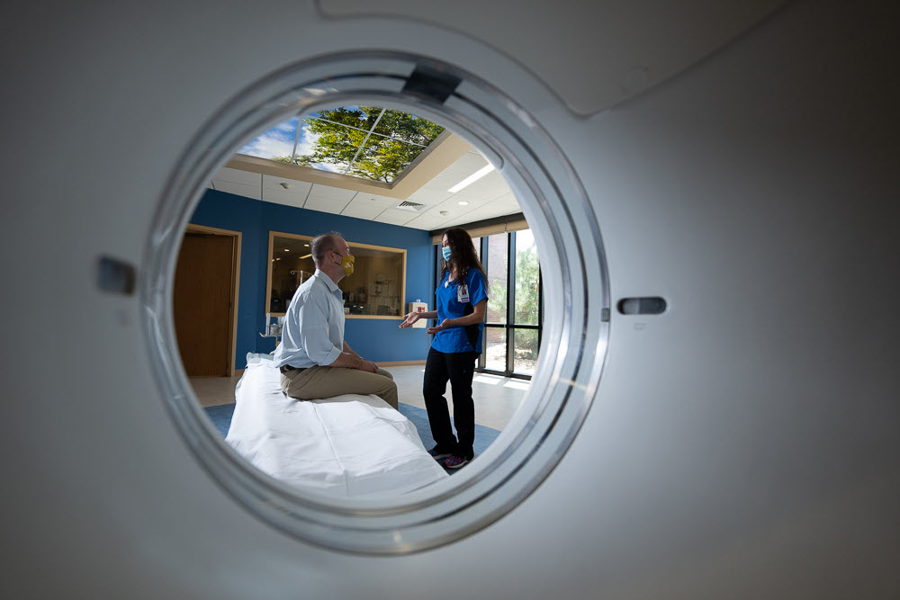
Before your imaging procedure, we’ll explain the test and answer any questions you may have. After your imaging procedure, a radiologist provides your doctor with interpretations of the findings. Radiologists are highly trained and experienced with reading the images and making assessments based on them. They then communicate their findings through highly detailed reports to your doctor, who will review the results with you. CommonSpirit imaging facilities are accredited in many procedures to ensure high-quality care, including MRIs, ultrasound, computed tomography (CT) scans, mammography, nuclear medicine, stereotactic breast biopsy and more. Accreditation is recognition for consistently meeting or exceeding high standards of care.
Many imaging services are non-invasive and don’t cause any pain. For more invasive procedures, our nurses and staff work with you to help make you comfortable and provide conscious sedation if necessary. Our specially trained nurses are certified in advanced lifesaving techniques. Ultimately, you and your family are our priority.
Most diagnostic images are taken digitally, enabling us to quickly and securely share your files with doctors anywhere in our network. Digital imaging can help your doctor provide you with an accurate diagnosis and begin treatment sooner. And if you’d like copies for your records, images can be saved to a compact disc or emailed anywhere in the world.
Our imaging centers offer a variety of services, but vary by location. Please confirm services directly with each location.
- Angiography: Angiography uses dyes and X-rays to take images of the blood flow in arteries or veins.
- Colonoscopy: Using a camera on a long, flexible tube, an examiner can screen or take images of the intestine linings. Learn more about how a colonoscopy can benefit you.
- Computed Tomography (CT) scans: CT scans merge multiple X-rays taken from different angles. The resulting cross-sectional images help doctors diagnose health problems of the bones and soft tissues.
- Dual-Energy X-ray Absorptiometry (DEXA) Scan: A DEXA scan is a specialized X-ray to determine whether you’re losing bone density in key areas, such as the hips and spine. It can help assess your bone strength and risk of fractures.
- Fluoroscopy: This imaging technology provides real-time moving images of your internal structures using a fluoroscope, which uses continuous X-rays to project images onto a screen. Fluoroscopic navigation can be used during surgery help surgeons actively track instruments during surgery to increase their precision.
- Interventional Radiology: Using CT scans, ultrasound and fluoroscopy to see inside the body, doctors use catheters or other small devices to provide diagnosis or treatment to open blockages in arteries, destroy tumors, treat aneurysms and more.
- Magnetic Resonance imaging (MRI): Using a strong magnetic field and radio waves, detailed images of the body and organs are created.
- Mammography: Using low-dose X-rays or ultrasound, doctors create a 3-D image of your breast to detect irregularities. Learn more about mammograms.
- Nuclear Medicine: Nuclear medicine is a branch of imaging that uses safe levels of radioactive material to diagnose or treat a wide variety of diseases and abnormalities.
- Positron Emission Tomography (PET) Scan: PET scans are a type of nuclear medicine imaging. Using a special dye, they measure organ and tissue functions such as blood flow, oxygen use and metabolism.
- Stereotactic Biopsy: Images are used to locate and target areas of concern, such as suspected tumors, to guide a biopsy. This helps ensure the doctor can take a sample of the right tissue to send to a lab for examination.
- Ultrasound: This type of scan uses high-frequency sound waves to produce images of soft tissues inside the body. Ultrasounds use no radiation and have a wide variety of applications, from detecting tumors and heart conditions to safely viewing a fetus during pregnancy.
- Intravascular Ultrasound (IVUS)/Fractional Flow Reserve (FFR): Using a catheter, IVUS/FFR enables real-time images of blood vessels to be taken from within the body to diagnose blockages.
- X-Ray: X-rays are a very common form of imaging. Small amounts of radiation are used to create images of bones and tissues inside the body. Our advanced equipment can reduce the amount of radiation required in these procedures
Advanced imaging technologies to enable you and your health care team get a better picture of your health. You can find imaging services at these locations near you.


